Digital pathology
Accelerate the path from images to answers

See how digital delivers.
For pathologists. For patients.
From swift slide scanning to confident diagnosis, our end-to-end solution unlocks collaboration and clinical depth, to find answers fast.
Philips Digital Pathology solution accelerates the path from images to answers, harnessing AI* to power faster, confident decision-making helping you deliver better care for more people.
Featured products in Digital pathology
-
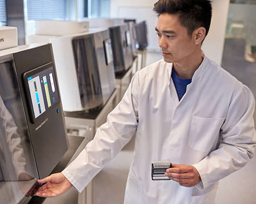
Pathology Scanner SG60
SG60 is designed to accommodate laboratories with a lean workflow and need to scan small batches of slides to achieve operational excellence and short turnaround times by scanning batches in parallel. With a high throughput, high first time right rate and load and walk away scanning, the SG60 enables you to digitize your histology samples and obtain high quality clinical diagnostic images for routine use and integrated pathology networks. Pathology Scanner SGi* with native configurable DICOM JPEG and JPEG XL output helps reduce file sizes without compromising diagnostic quality, unlocking significant storage savings and performance improvements.
FDP0909 -
Pathology Scanner SG300
SG300 is designed to accommodate laboratories for high volume labs that want to maximize scanner utilization and further reduce the total cost of ownership per slide by means of overnight scanning. With a high throughput, high first time right rate and load and walk away scanning, the SG300 enables you to digitize your histology samples and obtain high quality clinical diagnostic images for routine use and integrated pathology networks. Pathology Scanner SGi* with native configurable DICOM JPEG and JPEG XL output helps reduce file sizes without compromising diagnostic quality, unlocking significant storage savings and performance improvements.
FDP0911 -
Image Management System
Consisting of a pathology viewer and a server and storage application, Image Management System (IMS) is designed to enhance the efficiency and effectiveness of your pathology lab. The open, scalable design and new user interface integrate into your workflow and IT infrastructure environment. The IMS manages image repositories – including whole slide images (WSI) and gross images – and comprehensive LIS integration, networking, security, audit trail and archiving capabilities. Easy access to information and resources can enhance digital case review.
FDP0912


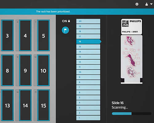
Fast scans and case review
If you leave the microscope behind, would you ever go back? Not many would. In a survey of 52 pathologists, 100% of them said that going digital helps reach diagnostic consensus and that they would never go back to non-digital.1 With our pathology slide scanners, the Image Management System (IMS) and a comprehensive set of software tools and capabilities, it’s easy to minimize doubt and aid confident diagnosis – fast.

What can Philips digital pathology do for you?
Our digital pathology solution enables more efficient, remote and collaborative ways of working. Pathologists have seen efficiency gains of up to 15-20% per case from making the digital transition.2 Going digital also helps reach diagnostic consensus.3 Unlocking expert second opinions drives improved diagnostic confidence so you can support making the right decisions for patients.

Open and secure image sharing
Our digital pathology solution can be used alongside with enterprise-wide IT infrastructure and AI capabilities*, allowing for cost-effective productivity and efficiency gains. For example, 35 pathology labs already use the Philips pathology solution and Ibex AI. This has been shown to result in up to 37% productivity gains for diagnosis.4
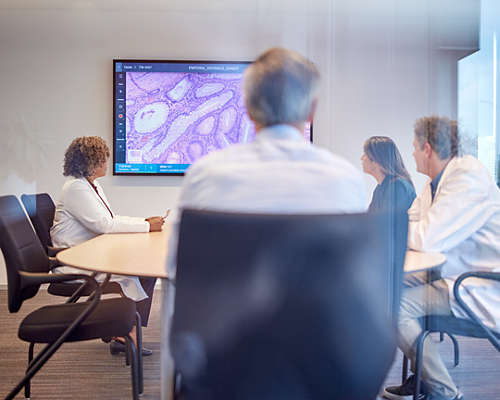
Confidence through collaboration
See what close to 400 customers and over 20 hospital pathology laboratories have learned about going fully digital with the Philips solution. It facilitates collaboration with clinical teams through remote consultation, real-time case sharing and multidisciplinary discussions.
Accelerate to digital
If you leave the microscope behind, would you ever go back? Not many would. In a survey of 52 pathologists, 100% of them said that going digital helps reach diagnostic consensus and that they would never go back to non-digital.1 With our pathology slide scanners, the Image Management System (IMS) and a comprehensive set of software tools and capabilities, it’s easy to minimize doubt and aid accurate diagnosis – fast.

See a return on investment (ROI) in just two years
Philips Capital offers a range of financial models to choose from, so you can benefit from improved diagnostic accuracy and optimized cashflow predictability. Our experts are ready to provide a custom consultation for your business and can even help calculate your projected ROI with Philips systems.

Healthcare IT: the nuances of integration & interoperability
Pathology is just embarking on its digitization journey, and there are multiple ways that departments can promote interoperability in partnership with industry. Standardizing whole slide image formats and promoting tight integration between different software systems will benefit not just individual users but also healthcare systems.
Technology and innovations
-
See how the Philips open platform approach for digital pathology helps achieve scanner integration with vendor-agnostic interoperability and open AI strategy.

Suites of solutions
-
Learn how Philips integrated diagnostics makes insights easily accessible across the enterprise, fostering collaboration and helping you accelerate diagnosis.
-
![Philips Healthcare for clarity on the cancer care journey]()
Philips helps bring more clarity at every moment of cancer care through streamlined multidisciplinary workflows and integrated patient data. See how.

Related stories


Results are specific to the institution where they were obtained and may not reflect the results achievable at other institutions
Footnotes
[1] Survey of 52 pathologists, lab managers and lab technicians in Europe, 2018. PIPS can be used for in vitro diagnostic purposes. The system can aid pathologists to review and interpret digital images of surgical pathology slides prepared from formalin-fixed paraffin embedded (FFPE) tissue. PIPS is not available for sale in all countries.
[2} KLAS Research ‘US Digital Pathology 2023’ Performance Insights.
[3]Sarfati D, Gurney J. Preventing cancer: the only way forward. The Lancet, August 2022, 400(10352):540-541. DOI: 10.1016/S0140-6736(22)01430-1.
[4] Information provided by Ibex AI Raoux, et al. Modern Pathology (2021) 34 (suppl 2): 598-599. Ibex AI is Research Use Only (RUO) in the US.
*PIPS enables iSyntax files and with the Software Development Kit (SDK) third-party companies can use this for AI capabilities.